
This illustration shows a technique described by Romain Gérard, MD, Richard Stern, MD, and Mathieu Assal, MD from the Orthopaedic Surgery Service, University Hospital of Geneva, Switzerland for the March 2008 issue of Orthopedics. This technique is valuable for providing a powerful and durable correction of metatarsus primus varus and hallux valgus, and careful attention to the details should help in achieving a successful outcome while avoiding complications.
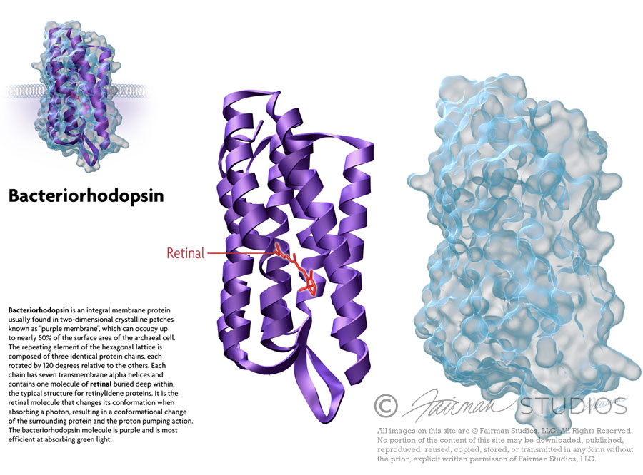
Bacteriorhodopsin is a protein used by Archaea, the most notable one being Halobacteria. It acts as a proton pump; that is, it captures light energy and uses it to move protons across the membrane out of the cell.[1] The resulting proton gradient is subsequently converted into chemical energy.[2]
Bacteriorhodopsin is an integral membrane protein usually found in two-dimensional crystalline patches known as “purple membrane”, which can occupy up to nearly 50% of the surface area of the archaeal cell. The repeating element of the hexagonal lattice is composed of three identical protein chains, each rotated by 120 degrees relative to the others. Each chain has seven transmembrane alpha helices and contains one molecule of retinal buried deep within, the typical structure for retinylidene proteins.
It is the retinal molecule that changes its conformation when absorbing a photon, resulting in a conformational change of the surrounding protein and the proton pumping action.[3] It is covalently linked to Lys216 in the chromophore by Schiff base action. After photoisomerization of the retinal molecule, Asp85 becomes a proton acceptor of the donor proton from the retinal molecule. This releases a proton from a “holding site” into the extracellular side (EC) of the membrane. Reprotonation of the retinal molecule by Asp96 restores its original isomerized form. This results in a second proton being released to the EC side. Asp85 releases its proton into the “holding site,” where a new cycle may begin.
The bacteriorhodopsin molecule is purple and is most efficient at absorbing green light (wavelength 500-650 nm, with the absorption maximum at 568 nm).
Bacteriorhodopsin belongs to a family of bacterial proteins related to vertebrate rhodopsins, the pigments that sense light in the retina. Rhodopsins also contain retinal; however, the functions of rhodopsin and bacteriorhodopsin are different, and there is only slight homology in their amino acid sequences. Both rhodopsin and bacteriorhodopsin belong to the 7TM receptor family of proteins, but rhodopsin is a G protein-coupled receptor and bacteriorhodopsin is not. In the first use of electron crystallography to obtain an atomic-level protein structure, the structure of bacteriorhodopsin was resolved in 1990. It was then used as a template to build models of G protein-coupled receptors before crystallographic structures were also available for these proteins.
Many molecules have homology to bacteriorhodopsin, including the light-driven chloride pump halorhodopsin (for which the crystal structure is also known), and some directly light-activated channels like channelrhodopsin.
All other photosynthetic systems in bacteria, algae, and plants use chlorophylls or bacteriochlorophylls rather than bacteriorhodopsin. These also produce a proton gradient, but in a quite different and more indirect way involving an electron transfer chain consisting of several other proteins. Furthermore, chlorophylls are aided in capturing light energy by other pigments known as “antennas”; these are not present in bacteriorhodopsin-based systems. Last, chlorophyll-based photosynthesis is coupled to carbon fixation (the incorporation of carbon dioxide into larger organic molecules); this is not true for bacteriorhodopsin-based system. Thus, it is likely that photosynthesis independently evolved at least twice, once in bacteria and once in archaea.
Sources:

The purpose of this illustration is to represent the anatomical systems of the body attacked by S. Pneumoniae, a bacteria which causes various diseases and infections, especially in young children of Third-World countries. The systems portrayed are the respiratory, nasopharyngeal, and circulatory systems. These systems are easily infected by this bacteria, causing illnesses such as pneumonia and sepsis. This image is used for public awareness and education.
Title: Systems of Pneumococcal Infection
Medium: Graphite Pencil, Adobe Photoshop
Format: Poster or PR presentation
Client: PneumoADIP, JHU Bloomberg School of Public Health
Audience: General public

This illustration was created for an article by Rita Rubin in USA Today entitled “Natural orifice’ surgery has tongues wagging.”
Surgeon Lee Swanstrom of the Oregon Clinic removed a patient’s gallbladder through her mouth. Here’s how:
1. An endoscope was snaked through the patient’s esophagus and into her stomach.
2. Next, a tiny knife was inserted through the scope and cut a quarter-inch to three-eighths-inch hole in her stomach. He then passed a tiny balloon through the scope and inflated it when it got to the end, stretching the hole.*
3. The surgeon dissected the patient’s gallbladder and pulled it back through her stomach and out her esophagus. (At 4 or 5 inches long, it was too large to bring through the scope and out of her mouth in one piece.)
4. Finally, the hole was closed in the patient’s stomach with sutures.
* Note: The surgeon also made two tiny incisions in her abdomen through which he could monitor what was going on. The goal, though, is surgery with no abdominal incisions.
Source: USA TODAY research

The mission of Space Medicine Associates is to provide multidisciplinary space medicine and bioastronautics consultation, training and oversight in support of all aspects of off-world travel. Serving both the commercial launch industry and the personal space traveler, SMA endeavors to safely open human spaceflight to as many individuals as possible.
SMA was founded in 2007 by four individuals of varied healthcare backgrounds – aerospace medicine, nursing, and pharmacy – who share the common vision of safely opening human spaceflight to as many individuals as possible. Thus the four stars in the final logo were used to represent the 4 founders.


 Loading ...


